COSMETIC PTERYGIUM SURGERY
If you value your eyes choose Dr. Khanna because:
– He has Pterygium surgery experience of close to 3 decades
– Has been successful with multiple procedures for over 25 years
– Uses the current, best, cosmetic strategy with the least chance of recurrence
– # 1 option of movie stars, newsreaders and medical professionals
– Board Licensed by American Board of Ophthalmology
– Pain-free surgical treatment in operating suite
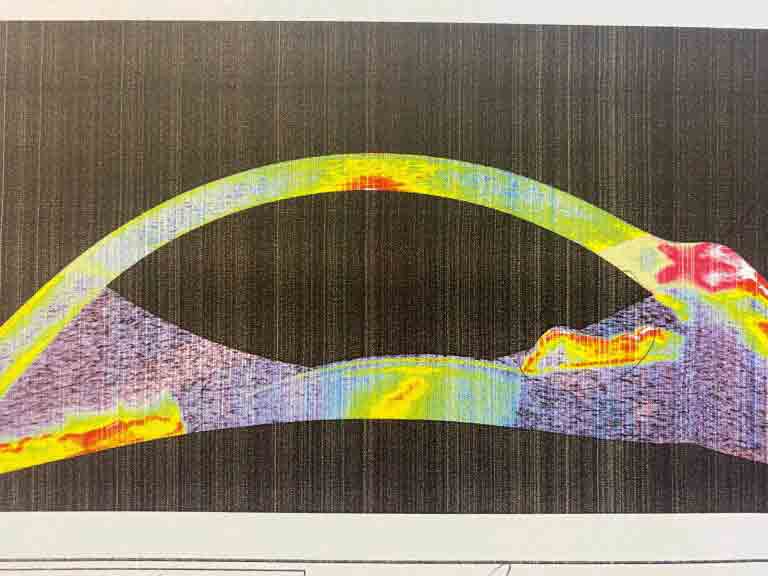
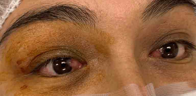
Elastotic subhyaloid degeneration of the conjunctiva.
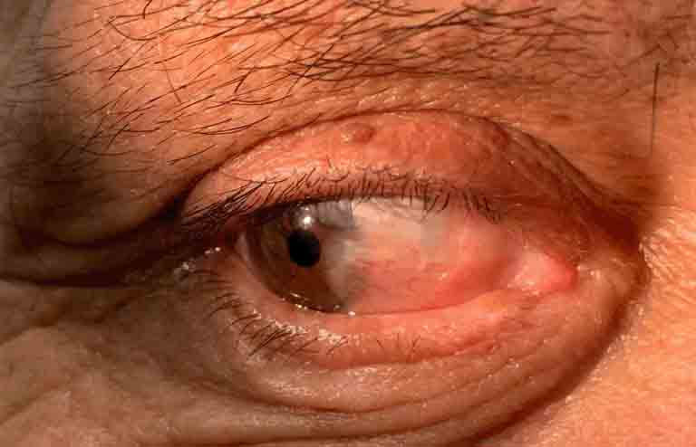
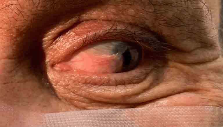
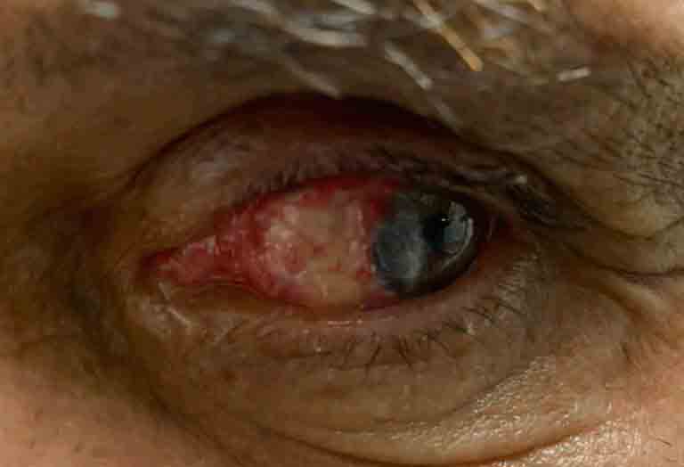
What is Pterygium or Surfer’s Eye?
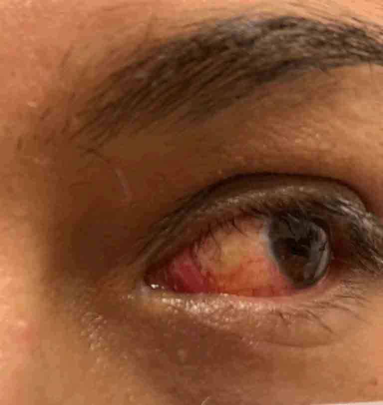
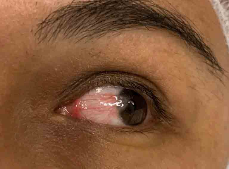
A pterygium is a light red or pinkish, elevated, triangular growth of tissue on the eyeball that starts on the white of the eye, also known as the sclera. This tissue growth can get into the cornea, which is the external, clear layer of the eye, where light enters the eye. Pterygium typically begins on the cornea near the nose. It could also gradually increase to a bigger size that may cover the pupil. If someone has more than one of these eye growths, the plural is pterygia. Pterygia are usually, benign (non-cancerous) growths, nonetheless, they can come to be malignant.

Basic synonyms:
Though it’s generally called ” surfer’s eye,” you do not require to be a surfer, or ever have seen the sea, to have a pterygium. However, being in extreme sunlight for lengthy hours – especially when get in the water, which reflects the sunshine’s dangerous UV rays – increases your risk.
Pingecula
An elevated, yellowish nodular growth on the conjunctiva. It is non-cancerous, however cosmetically unsightly. Its etiology, prevention as well as treatment are similar to pterygia. In fact, it might be considered a beginning of a pterygium.
Causes of Pterygium progression
Although ultraviolet radiation from sunshine appears the primary reason for the growth of pterygia, dirt, in addition to wind, are also connected in some cases. Another factor for the growth is dry eye disease. Pterygia generally develops in 30 to 50-year-olds. These bumps on the eyeball are hardly ever seen in youngsters. Having light skin, along with light eyes, might place you at increased risk of getting a pterygium.
Symptoms of Pterygium
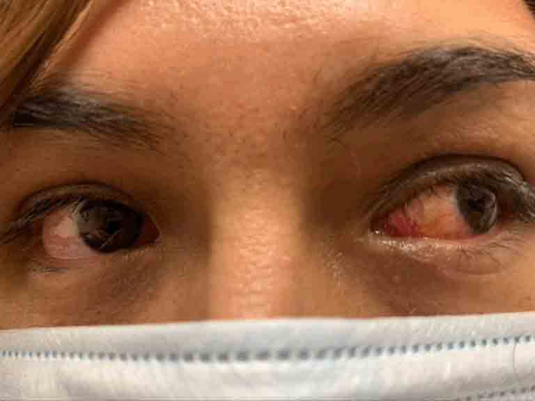
Pterygium is a callus like growth on the clear part of the eye (the cornea). It can be undesirable and people dislike its appearance. A lot of people wrongly assume the person is drunk. It can make life difficult at the office, particularly if you are making a presentation to close a big deal. You might have spent a large amount of time in front of the mirror questioning why your eyes always turn red, or taking a look at a growth covering the white part of your eye. Pingecula causes similar symptoms.
Prevention of Pterygium
There are 2 ways to stop onset or progression of Pterygium:
1. Avoid UV radiation
Use wide brimmed hats as well as sunglasses. If you are a surfer, try tinted swimming goggles. Decreasing UV radiation prevents degeneration of conjunctiva.
2. Treat dry eyes
Preservative free artificial tears, ointment, medicated drops etc. Find out more about dry eyes.
Pingecula or Pterygium Drops
Drops are a very important part in our fight against pingecula developing into a pterygium, progression of pterygium and the prevention of recurrence. At 1800realdoctors.com, our team believes surgery might to be the last resource. We are happy to state we have actually prevented operations in several patients by cautious use of the following drops and accessories:
Water droplets: The simplest way to moisturize the eye is to sprinkle them with water. A humidifier is a cost effective, excellent way to keep the eyes moist gently. Washing your eyes by dipping them in a cup full of cool, filtered water serves to moisturize them, along with ridding them of trapped allergens.
Artificial tears: One of the most popular techniques in America. Chemical free, artificial tears of various brand names can be found in single use, plastic containers. They are better than multidose bottles which have preservatives and may be toxic to cornea.
Ointments: A lot of pharmaceutical companies that make artificial tears also make eye ointments. These are better than drops due to the fact that they need usage only at nighttime. They stick to the cornea and coat it. Only a little amount must be used at a time.
Ayurvedic drops: For centuries, these have proven valuable in the Indian subcontinent and beyond. Gulab Jal is made from rose petals. We provide freshly made drops from organically grown roses.
Medicated drops: Restasis was the very first to be introduced, followed by the quicker acting Xiidra. Now, Cequia has been introduced. These are all available by prescription only.
Solid drops: A strong, small cylindrical rod is inserted between the lid and the eye by the patient. This material gradually liquifies, giving artificial tears.
Tear preservers: Small microscopic rods made from collagen or synthetic material are inserted in the drainage pathway of the tears by an accomplished doctor in a quick procedure in the office. These preserve the natural tears which have properties to fight the growth of pingecula.
Anti-cancer medicine: Mitomycin C is an anticancer agent which has been shown to shrink pterygium. It can be used as drops or the doctor can inject it under the pterygium, especially recurring ones.
Symptoms for Pterygium Surgery:
There are two important considerations to remember when thinking of getting your pterygium removed. If it starts to grow, it needs to be removed before it causes long-term visual changes. Secondly, meticulous surgery by a skilled pterygium doctor is needed to get great cosmetic results and also prevent recurrence.
1. Visual:
Causes astigmatism, affecting vision.
Invades the center of the cornea, preventing light to enter into the eye
Restriction of eye movement
Dry eyes, because of interference in the spread of tears
2. Cosmetic:
High schooler or College going person may find social interaction challenging
At work, people usually think a person with surfer’s eye is drunk
Contraindications:
Being on blood thinners or having an active bleeding condition
Distinguish from Pesudopteygium
Psudo-pterygium is a condition where conjunctiva covers an inflamed portion of the cornea. Pterygium can be differentiated from psudo-pteygium by doing a probe test. In a pterygium, a probe can be passed between the pterygium and the eyeball.
Before Treatment:
– You should stop taking blood thinners, like aspirin or warfarin
– Control dry eyes with preservative free artificial tears
– Start antibiotic and steroid drops or ointments
Day of Procedure:
– Relax, take an anxiolytic (anxiety reducing medication)
– Bring along your favorite songs to listen to during the procedure
Surgery Steps:
– Mark out the entire pterygium and excise it
– Remove the pterygium totally from the cornea. Polish the remaining fibers to leave a clear surface
– Micro-dissect the structure on which the pterygium grows
– Avoid excessive blood loss which can cause long term redness and swelling. Cauterize all bleeders
– Cover the exposed area where the pterygium was removed with conjunctival autograft or amino-graft allograft using tissue or other bio glue. This is a very important step. Previously, stitches were used to close the wound, however they can induce inflammation and pain. Tisseel glue is self-dissolving and pain-free, so it produces more cosmesis.
After the Operation:
– Taper the steroid and non-steroid drops as opposed to stopping them abruptly
– Use punctal occluders, if necessary. Keep the eye hydrated and moist
– Watch out for early recurrence
– Treat early recurrence with anti-inflammatory drops or Mitomycin injection
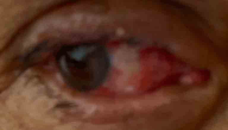
Recurrent Pterygium Management
Measure 10 times and operate once – planning is the key!
Persistent pterygium may be considered a typical adverse effect of pterygium surgery. If the pterygium is removed, however, no attention is paid to removing the structure of tenon’s capsule, nor are adjunct treatments used, there is up to a 70% possibility of recurrence. Recurrent pterygium management is an art and science. We end up helping a lot of these patients who had their first pterygium surgery in other places. Pterygium are like weeds, either behead them, kill them entirely or leave them alone. Never ever prick or irritate them because then they attack savagely.
Reoccurring pterygium is worse than the initial one due to the fact that it can spread in all directions and have extra fibrosis. This means they are firmly affixed to the muscle and surrounding eye tissue. They are also a lot more vascular, because blood vessels provide it the nourishment it requires in its march to conquer territory it had lost previously.
Video: Is your pterygium back once again? The above video tells you important information about what you need to know about recurring pterygium and pterygium removal
Before operating on a recurring pterygium:
– Find out what was done in the original procedure
– Treat dryness of the eyes
– Plan to repeat surgery in winter or when the UV exposure is going to be minimum
– Inject Mitomycin C (avoid in pregnant or breast-feeding women) into the pterygium body. This anti-cancer medicine kills the cells of the pterygium, including the blood vessel cells. This causes the recurring pterygium to shrink in size and its margins to become more defined.
– The surgical treatment might be more excruciating, so attention should be directed towards proper numbing of the eye and oral or IV pain relievers
During the procedure of recurrent pterygium:
– Identify the borders and extensions of the recurrent pterygium
– Identify the muscles like the lateral rectus, medial rectus and the obliques and be sure to avoid injury to them
– If conjunctival autograft does not suffice, an allograft might be required
– Attention to good control over bleeding is necessary
– Avoid using stitches
After surgery for recurrent pterygium:
– Close follow up is needed
– Treat emerging blood vessels aggressively
– Taper anti-inflammatory drops gradually
Please don’t worry if you have had a recurrence of pterygium. If you operate out of the Los Angeles area, you can FaceTime, Skype or Zoom with us to find out about your options. If you send us your insurance cover card, our staff can let you know your insurance coverage.
Rajesh Khanna, MD has been carrying out pterygium treatment for almost thirty years. Over the decades, he has made use of numerous, prevalent techniques of pterygium surgery. Obviously, he has helped hundreds of people across the globe seeking relief. Now, he has perfected the art of a cosmetic outcome with a really reduced possibility of recurrence.
Link: Find out more about our Pterygium specialist
Pterygium and Lasik Eye Surgery
Pterygium, which is a callus growth on the eye, is a result of dry eye. Lasik can even increase dryness of the eyes. Hence, Lasik eye surgery may cause progression of pterygium. Depending upon the decision of the surgeon, Lasik eye surgery may need to be avoided in the presence of pterygium.
There numerous reasons why Lasik may be deemed unsafe if you have Pterygium:
– Through the process of making the Lasik flap, the head of the pterygium may get cut. This is a big problem for the patient since an irritated pterygium is even worse for the eye
– The pterygium may grow into the interface, causing visual problems. Because the potential space permits it an unobstructed progress, it might expand in all directions while lifting the flap. This results in irregular astigmatism, causing ghosting of images.
– The progressive pterygium might grow over the flap. Removing it might be extremely difficult, as it might lift the flap
– The pterygium may grow over and under the flap and this will affect the nutrition of the flap, leading to the destruction of the edges of the flap
So, what are the options to see better in the presence of pterygium?
– Superlasik eye surgery is a wonderful choice for people suffering with pterygium
– An additional alternative is Visian ICL (Implantable Collamer Lenses) inserted away from the pterygium.
– The most effective option is Superlasik eye surgery combined with pterygium removal. Superlasik, as you might recall, uses a highly specialized automated device called epi-keratome. This divides the top layer of the cornea along a natural cleavage plane. The same plane is needed to separate the progressive pterygium.
Therefore, combining the two procedures makes a lot of sense. It does require advanced instrumentation and higher skill; however, this is likely the best option to deal with two problems simultaneously. Also, as both treatments require similar medications it saves money and time for the eye drops and it will decrease the total number of visits to the Lasik eye surgeon.
VIRGIN EYES (WITH INSURANCE COVERAGE)
Only PPO accepted. With HMO you will need to coordinate care with your HMO physician.
$.49.99/mo *
– Pre-operative work up
– Free Tea & Coffee
– Clinic costs
– Glue and grafti
– Punctal occluders
– Please email insurance card
VERIFY NOW
VIRGIN EYES (No Insurance Coverage)
One of the most popular choices
$99.99/mo *
– Pre-operative work up
– Free Tea & Coffee
– Clinic fees
– Glue and grafti
– Punctal occluders
– 12 month follow up
BOOK NOW
RECURRENT PTERYGIUM
Simple, fast and effective flexible move
$149.99/ mo *
– Pre-operative work up
– Free Tea & Coffee
– Clinic costs
– Glue and grafti
– External grafti
– Mitomycin injection
– Punctal occluders
– twelve month follow up